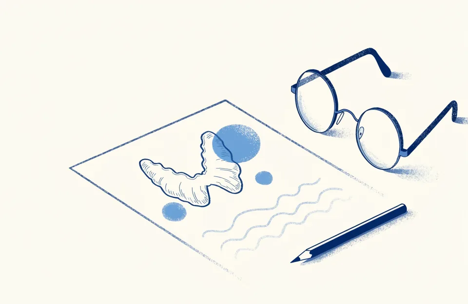
- The report describes five features of the nodule: what it is made of, how dark it looks, its shape, its edges and whether it has bright spots.
- The diagnosis does not come from a single word but from all of them together: the radiologist scores each feature, and the total gives the TIRADS category, which decides whether a biopsy is needed.
- The volume in milliliters matters more than most people realize, because it affects which treatments are an option.
Most patients arrive at the consultation having read the report several times and understood only part of it. That is normal: it is written for another doctor, not for you. Here are the terms that come up most often, in plain language and in the order they usually appear.
What the nodule is made of
A nodule can be A nodule made of tissue, with no fluid inside. It is the most common type., A nodule filled with fluid. It is usually benign and, if it causes symptoms, is treated with its own technique. or mixed. A solid nodule is made of tissue, a cystic one is filled with fluid, and a mixed one has both. Those that are mostly fluid are less worrying and, if they cause symptoms, are treated with a different technique than solid ones. You may also see the word A nodule with many small spaces, like a sponge. It is considered benign with a high degree of confidence.: a nodule full of tiny spaces, like a sponge, which is considered benign with a high degree of confidence.
How dark it looks
The shade of gray a tissue shows on ultrasound, compared with its surroundings. is how gray the nodule looks compared with the surrounding tissue. Darker than normal thyroid tissue. It adds two points to the TIRADS score, and on its own it does not mean cancer. means darker, isoechoic means the same shade, and hyperechoic means lighter. A darker nodule scores a little higher, but on its own that means little: many benign nodules are hypoechoic.
Shape and edges
There are two phrases that do deserve attention. “Taller than wide” describes a nodule growing against the natural direction of the tissue, and it adds points. “Irregular” or “lobulated margins” describes edges that are not well defined, and that adds points too. The opposite, “well defined” and “wider than tall”, is what most nodules look like.
The bright spots
Very small, bright dots inside the nodule. They are one of the features that carry the most weight in the TIRADS score. are very small, bright dots inside the nodule, and they are among the features that carry the most weight in the score. Large or “eggshell” calcifications weigh less. And there is one artifact that is reassuring: the A bright trail behind a dot. It is associated with colloid content, that is, with a benign nodule., a bright trail behind a dot, which is associated with benign nodules containing colloid.
How it all adds up: the TIRADS score
The radiologist takes those five features, scores them and obtains a category from 1 to 5. That is your TIRADS category. It is not a diagnosis but a probability, and it determines whether a biopsy is needed and at what size. A small TR3 is monitored, and a TR5 is studied further. The categories and their thresholds are explained on the nodules page.
The number almost nobody looks at: the volume
The report usually gives three measurements in millimeters and, sometimes, the volume in milliliters. That number matters for two reasons. First, because growth is measured by comparing volumes between one ultrasound and the next, not by comparing the largest diameter. Second, because it affects which treatments are an option: when a benign nodule is treated without surgery, larger nodules shrink less and sometimes need more than one session.
- 20 mLis the volume above which, in the literature, ablation shrinks the nodule less: success of 64.5% versus 81 to 87% in smaller ones.Korean Society of Thyroid Radiology (KSThR) guideline, 2017. It is a predictor of response, not an exclusion.
- Tessler FN et al. ACR Thyroid Imaging, Reporting and Data System (TI-RADS). J Am Coll Radiol. 2017, 14(5): 587-595.
- Kim JH et al. 2017 Thyroid Radiofrequency Ablation Guideline: Korean Society of Thyroid Radiology. Korean J Radiol. 2018, 19(4): 632-655.
